
👉👉 🇺🇸 All Posts 🇬🇧 / 🇯🇵 記事一覧 🇯🇵 👈👈
♦️ Introduction
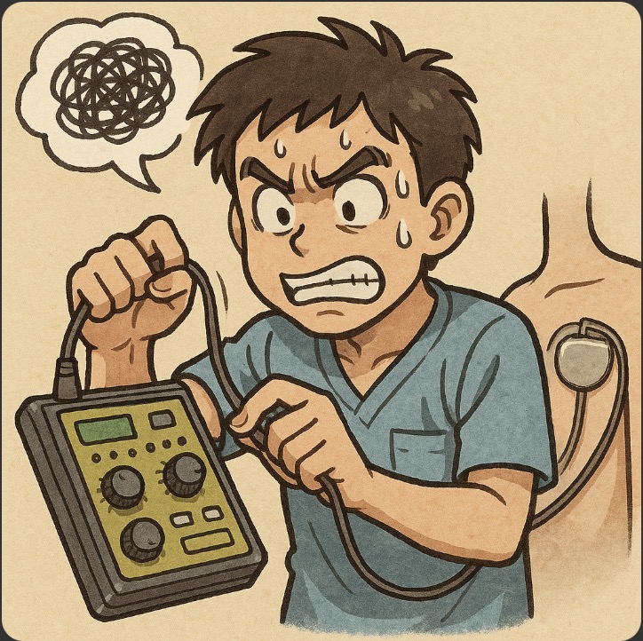
If you’ve worked in the OR or ICU, you’ve probably seen a temporary pacemaker placed via the internal jugular vein with that mysterious dial-box generator dangling at the bedside 😅.
Before surgery, someone may place a programmer head over the chest (right above the implanted device) and use a computer-like programmer to tweak settings—many centers now also use remote management.
In cardiac surgery, you’ll see epicardial wires sewn onto the myocardium and “handed over” to anesthesia to manage pacing from our side—classic cardiac OR “hand-off.”
Pacemakers (like ECG interpretation) can feel intimidating, so they are frequent exam topics in oral/written anesthesiology boards. Let’s lock in the basics.
Besides standard pacemakers, you’ll encounter ICDs (implantable cardioverter-defibrillators) and CRT-D (cardiac resynchronization + defibrillator). We’ll start with the basic pacemaker first.
♦️ Why implant a pacemaker?
Textbooks list many indications, but the core idea is simple: symptomatic bradycardia with inadequate cardiac output. Typical examples:
- Sick sinus syndrome (SSS)
- High-grade AV block (Mobitz II, complete/third-degree)
- Bradycardic atrial fibrillation
- Post-MI low-output states
- Temporary support after cardiac surgery (epicardial pacing)
(For fine details, defer to cardiology texts/guidelines—no one memorizes every line item 😉.)
♦️ NBG Pacemaker Code (the “mode” code)
Saying “Back-up VVI at 40” sounds slick—but don’t worry, that vibe is mostly imaginary 😎. If you grasp the first three letters, you can handle most conversations.
- 1 = Chamber paced,
- 2 = Chamber sensed,
- 3 = Response to sensing
- (4 = Rate response, 5 = Multisite pacing).
🔷 First letter (O / A / V / D)
The first letter indicates the chamber that is paced. Since the pacing site is the most essential feature, it comes first in the code.
- O = No pacing (in which case the pacemaker essentially has no function!)
- A = Atrium
- V = Ventricle
- D = Dual (both atrium and ventricle; note: “D” stands for dual, not double).
🔷 Second letter (O / A / V / D)
The second letter indicates the chamber where the patient’s intrinsic electrical activity is sensed. If the patient is trying to generate their own depolarization, the device should not pace over it; conversely, if no intrinsic activity is present, the pacemaker must deliver stimulation.
- The meanings of O, A, V, D are the same as for the first letter.
🔷 Third letter (O / I / T / D)
The third letter indicates how the pacemaker responds to the sensed activity (from the second position). Four options are used:
- O = No response to sensing. Importantly, this does not mean “no pacing.” It means that pacing occurs regardless of whether intrinsic activity is present or absent → i.e., asynchronous pacing. (This can, in some circumstances, trigger ventricular arrhythmias such as spike-on-T.)
- I = Inhibited: when intrinsic activity is sensed, pacing is withheld.
- T = Triggered: pacing is delivered in synchrony with the sensed activity.
- D = Dual: both inhibited and triggered functions apply.
The terminology can be confusing. For example, “triggered” means: if atrial activity is sensed but not conducted to the ventricle, the device can deliver ventricular pacing synchronized to the atrial event. If there is no atrial activity and the device paces the atrium, but this fails to conduct to the ventricle, it can deliver ventricular pacing synchronized to its own atrial output. In practice, the most commonly used settings are I (inhibited) or D (dual).
Note: The pacing output strength and the sensing threshold (what counts as “present” vs “absent” intrinsic activity) can be adjusted in the device programming.
🔷 Mini-summary
In daily practice, if you understand these first three letters, you can manage most situations.
The most frequently encountered modes are VVI and DDD. Less commonly, you may encounter VDD or DDI, while AAI is rarely used.
🔷 Fourth letter (O / R)
The fourth letter indicates the presence of a rate modulation (rate response) function. This allows the pacemaker to increase heart rate in response to physiologic demand, by sensing activity such as motion, temperature, or minute ventilation.
- O = No rate response
- R = Rate response present
For example, you may have seen devices programmed as DDDR.
Patients with intact chronotropic competence (able to increase their own heart rate) may do fine without this feature. But in pacemaker-dependent patients, the absence of rate response can mean they become presyncopal after only minimal exertion, since their HR cannot rise appropriately.
🔷 Fifth letter (O / A / V / D)
The fifth letter indicates multisite pacing:
- O = None
- A = Multiple atrial sites
- V = Multiple ventricular sites
- D = Dual (e.g., both atrial and ventricular multisite pacing)
This is mainly relevant in cardiac resynchronization therapy (CRT), where biatrial or biventricular pacing is used to improve synchrony.
♦️ Preoperative Checklist (what to verify)
Start with the device card.
Confirm below.
- indication,
- current mode,
- battery status,
- last interrogation date.
- If the card is missing, call the managing clinic.
Look at imaging & ECG.
- Chest X-ray: generator position, lead number/course
- ECG: pacing spikes, morphology, and degree of device dependence
Coordinate with the device team (industry rep or clinical engineer). Ask for:
- Updated interrogation (typical intervals: ≤12 months for PM; 3–6 months for ICD/CRT-D)
- A perioperative plan (e.g., dependence → asynchronous; disable ICD therapies; post-op reprogramming)
- On-site support during the case if appropriate.
Plan the surgery and Bakup pacing ready:
- Will the surgeon use monopolar cautery or can they use bipolar/ultrasonic alternatives?
- Ensure transcutaneous or transvenous temporary pacing is immediately available.
♦️ Intraoperative settings & anesthesia
🔷 Anesthetic technique:
Use whatever fits the case. Turn OFF “pacing-spike suppression” filters on monitors so you can see spikes. Continuously confirm effective perfusing beats (SpO₂ waveform, arterial line, auscultation).
🔷 Why change device settings?
The main reason is to avoid electromagnetic interference (EMI). Devices/procedures to watch out for include electrosurgery, RFA (radiofrequency ablation), ESWL (extracorporeal shock wave lithotripsy), MRI (if the device is MR-conditional, then it may be acceptable under specified conditions), radiation therapy, and MEP/nerve stimulators. Here we will focus on electrosurgery, which we frequently use.
- Monopolar electrosurgery: the current flows from the pencil through the patient’s body to the dispersive (return) electrode, so the body is part of the current path.
- Bipolar electrosurgery: the current is confined between the tips of the forceps, completing the circuit locally.
What becomes problematic in pacemaker patients is the use of monopolar cautery. You have probably seen the ECG become messy when the cautery is activated; the pacemaker may misinterpret the cautery noise as intrinsic cardiac activity and, due to the I (inhibited) response, withhold pacing when it actually should pace.
If a patient has a strong intrinsic rhythm, that may be fine; but in a pacemaker-dependent patient, this can lead to severe bradycardia. (From the pacemaker’s point of view: “Hey, you’re beating on your own—I’m not needed,” so it stays quiet.)
To prevent this, preoperative programming changes are needed.
As an aside: in psychiatry, bipolar disorder refers to manic–depressive illness (two “poles,” mania and depression). So when psychiatrists say “bipolar,” they mean that, not the electrosurgical device—don’t mix them up 😅 (though you probably wouldn’t!).
🔷 If monopolar electrocautery must be used (specific strategies)
Use bipolar or ultrasonic devices whenever possible.
- Keep it short and intermittent (e.g., 1–2 seconds at a time, with sufficient intervals) and at the lowest possible power setting.
- Prefer CUT mode whenever possible. (COAG mode has been reported to cause strong EMI leading to syncope or even asystole).
- Place the dispersive (return) electrode close to the surgical field, but away from the pacemaker generator (at least ~15 cm), and position it so that the electrical current path does not cross the device.
- Have a defibrillator immediately available; apply defibrillation pads but avoid placing them over the device.
- Do not use cautery directly above the generator (within 10–15 cm), as this carries a risk of damaging the device itself.
Although more recent CIEDs have improved EMI resistance, situations involving close proximity, high output, or long-duration current flow still carry significant risk.
🔷 Basic intraoperative principles
There are many details, but the fundamental modifications can be summarized into three main categories:
- Turn off the rate-response function and ICD therapies immediately before surgery.
- Rate response: to avoid inappropriate tachycardia triggered by ventilator motion or changes in ventilation.
- ICD therapies: because cautery noise can be misinterpreted as ventricular fibrillation, resulting in inappropriate shocks.
- Decide between asynchronous pacing (AOO, VOO, DOO) or a mode that guarantees a minimum heart rate (AAI, VVI, DDI).
- The former is used for pacemaker-dependent patients when monopolar electrocautery is required. These patients cannot maintain an adequate heart rate on their own if the device is inhibited.
- The latter is for patients with relatively preserved intrinsic heart rate (≥50 bpm), where the pacemaker only needs to serve as a safety net if intrinsic activity fails.
- What is asynchronous pacing?
- As the name implies, it means pacing continues regardless of whether intrinsic cardiac activity is present or absent.
- The advantage: cautery noise is ignored, so the patient maintains a steady rate.
- Typically, the pacing rate is set slightly faster than the intrinsic rate, usually 80–90 bpm, to ensure capture during surgical stress.
For patients who still have sufficient intrinsic heart rate, asynchronous pacing can lead to pacing spikes falling on the T wave (spike-on-T) and provoke ventricular arrhythmias (rare in practice but theoretically possible).
In such patients, it is better to use a mode that provides a minimum guaranteed heart rate should their intrinsic rhythm fail—such as AAI, VVI, or DDI.
However, AAI is contraindicated if there is AV conduction disease or atrial fibrillation, since atrial pacing will not reliably conduct to the ventricles.
In patients who are minimally device-dependent, sometimes no programming change is made at all.
After surgery, always perform a device check and restore the original programming.
A note on modern devices
Recent CIEDs have indeed become more resistant to EMI. However, the combination of close proximity, high power, long-duration cautery, or poorly positioned current pathways remains a recipe for trouble.
The true “three essentials” are:
Preoperative planning + Intraoperative supervision + Postoperative reprogramming.
🔷 Key bullet reminders
- Turn OFF rate-response and ICD therapies (to prevent inappropriate tachycardia or shocks).
- Pacemaker-dependent: set asynchronous pacing (AOO/VOO/DOO) at a steady rate (typically 80–90/min).
- Not dependent (intrinsic rhythm adequate): use VVI/AAI/DDI for minimum backup (avoid AAI if AV block or atrial fibrillation).
- After surgery: always re-check and restore original settings.
At the end of the day, when you hear senior staff speak about pacemaker programming in the OR, it may sound like they are showing off—but in reality, it isn’t as complicated as it seems 😅. Don’t let yourself develop a “pacemaker allergy”!
♦️ Pacemaker malfunction (quick pattern recognition)
🔷 Pacemaker malfunction (quick overview)
Finally, a brief note on pacemaker malfunction. By definition, this means situations where the pacemaker does not function properly. Of course, we would prefer this never to happen; fortunately, in my own practice, I’ve never personally experienced this outside of cardiac surgery cases.
The causes of pacemaker malfunction can be broadly divided into two categories:
- Problems with output (and its effect)
- Problems with sensing
🔷 Output problems
Reasons why pacing output may not be delivered properly include:
- Electrocautery used very close to the generator, causing the device itself to fail (rare).
- Battery depletion (preventable if you check preoperatively).
- Lead fracture or dislodgement.
- Oversensing (see below), which suppresses output even though it is actually needed.
- Crosstalk: for example, atrial pacing output may be mistakenly sensed by the ventricular channel, leading the device to inhibit ventricular pacing.
Supplementary note:
When temporary epicardial pacing wires are sewn onto the heart in cardiac surgery, scar tissue may develop, requiring higher output or even repositioning of the wires.
So, in summary, output can fail because of:
- Generator failure (rare)
- Battery depletion (preventable with pre-op checks)
- Lead fracture/dislodgement
- Oversensing
- Crosstalk/misrouting
🔷 Sensing problems
Sensing abnormalities are generally divided into undersensing and oversensing.
Such inappropriate sensing often arises because cautery interference, antiarrhythmic drugs, myocardial ischemia, acid–base disorders, or electrolyte abnormalities can alter pacing thresholds.
- Undersensing = “a person who can’t read the room.” (Or more positively, “a person with the power of insensitivity.”) The pacemaker fails to notice that the heart is trying to generate intrinsic activity, and therefore delivers pacing inappropriately.
- Oversensing = “a person who worries too much about irrelevant things (hypersensitive).” The pacemaker misinterprets unrelated signals (e.g., cautery, muscle fasciculations with succinylcholine, EMG noise) as cardiac activity and therefore inhibits pacing inappropriately.
Personally, I used to be more of an “oversensing type” in life, but as I’ve gotten older, I think I’m gradually turning into an “undersensing type” human being… 😅
In brief:
- Undersensing = doesn’t notice intrinsic beats → paces inappropriately.
- Oversensing = misinterprets non-cardiac noise as intrinsic beats → pacing suppressed. (Either can be worsened by antiarrhythmics, ischemia, pH/electrolyte shifts.)
🔷 What to do if pacemaker malfunction leads to bradycardia…
If pacing failure occurs and the patient develops severe bradycardia, how should we respond?
- Magnet mode: If asynchronous pacing is not already enabled, placing a magnet over the generator can switch the device into magnet mode (asynchronous pacing), which may help in cases of oversensing. However—don’t just do this on your own! The magnet response differs by manufacturer:
- Most pacemakers → fixed-rate “magnet mode.”
- Most ICDs → only tachyarrhythmia therapies are disabled; pacing remains as programmed. → Always use magnets only as part of a planned strategy.
- Temporary pacing (transcutaneous or transvenous). This is the most straightforward option.
- Pharmacologic support:
- Use sympathomimetics (epinephrine, dopamine, isoproterenol) to lower the depolarization threshold and/or raise the heart rate.
- Correct electrolyte abnormalities (K⁺, Ca²⁺, Mg²⁺).
- Be aware that amiodarone increases the depolarization threshold; if the patient is on it, reduction or discontinuation may be required.
- Final fallback: direct epicardial pacing.
☝️ Practical checklist
- Do not use a magnet arbitrarily (responses differ by device).
- Prepare for temporary pacing quickly if needed.
- Sympathomimetics (epi/dopamine/isoproterenol) can help.
- Correct electrolytes, reconsider amiodarone.
- Last resort: direct epicardial pacing.
📝 Summary
☝️ Basics
- CIED (Cardiac Implantable Electronic Device) = Pacemaker, ICD, CRT-D.
- Main indication: symptomatic bradycardia with inadequate cardiac output.
- Examples: Sick sinus syndrome (SSS), high-grade AV block, bradycardic atrial fibrillation.
- NBG code:
- 1= chamber paced
- 2= chamber sensed
- 3= response to sensing ( I = inhibited, T= triggered, O = none)
- 4= rate response
- 5= multisite pacing
→ Most common modes: VVI, DDD.
☝️ Preoperative checks
- Always check the device ID card (indication, mode, battery status, last interrogation date).
- Chest X-ray: lead number and location.
- ECG: pacing spikes, morphology, degree of dependence.
- Consult device team (engineer/industry rep): interrogation, reprogramming if needed, intraop support, and postoperative restoration.
- Confirm surgical plan: monopolar vs bipolar/ultrasonic cautery.
- Prepare for backup pacing (transcutaneous or transvenous).
☝️ Intraoperative management
- Anesthetic technique: no special restrictions.
- Turn OFF “pacing spike suppression” on monitors.
- Continuously verify effective perfusing beats (SpO₂, arterial line, auscultation).
- Disable ICD therapies and rate response.
- If pacemaker-dependent → use asynchronous mode (AOO/VOO/DOO), typically 80–90/min.
- If not dependent → use minimal backup (VVI/AAI/DDI).
- Avoid AAI if AV block or atrial fibrillation.
- If the patient is barely dependent, sometimes no programming change is required.
- After surgery, always re-check and restore original settings.
- ☝️ Electrosurgery precautions
- Prefer bipolar or ultrasonic devices.
- If monopolar must be used:
- Short bursts, intermittent, lowest power.
- Prefer CUT mode (COAG mode causes stronger EMI; case reports of asystole).
- Place return electrode close to the surgical field, ≥15 cm away from generator, ensure current path does not cross the device.
- Have defibrillator ready; apply pads away from the device.
- Never use cautery directly over the generator.
- ☝️ Pacemaker malfunction
- Output problems: battery depletion, lead fracture/dislodgement, generator failure.
- Sensing problems:
- Undersensing = “can’t read the room” → fails to notice intrinsic activity → inappropriate pacing.
- Oversensing = “too sensitive” → mistakes unrelated noise (cautery, EMG, fasciculations) for intrinsic beats → inappropriate inhibition.
- Management:
- Magnets: device-dependent (pacemaker → fixed-rate mode; ICD → disables shocks but pacing unchanged). Use only as planned.
- Temporary pacing: transcutaneous or transvenous.
- Sympathomimetics (epinephrine, dopamine, isoproterenol).
- Correct electrolytes; consider withholding amiodarone if thresholds are high.
- Last resort: direct epicardial pacing.
📚 References & Links
- ASA Practice Advisory (2020) Perioperative Management of Patients with Cardiac Implantable Electronic Devices. Anesthesiology 2020;132:225–252. 👉 Practical steps for anesthesiologists: pre-op evaluation, intraop programming choices, post-op restoration. PDF available.
- HRS/ASA Expert Consensus (2011, reaffirmed later) Perioperative management of patients with implantable defibrillators, pacemakers and arrhythmia monitors. Heart Rhythm 2011. 👉 Foundational perioperative CIED guidance (magnet responses, electrosurgery patterns) referenced worldwide.
- EHRA Consensus (2022) Prevention and management of interference due to medical procedures in patients with CIEDs. Europace 2022. 👉 Europe-focused, procedure-specific EMI risk tables (MRI, RT, ESU, etc.) and peri-procedural monitoring advice; open access.
- AHA Scientific Statement (2024) Periprocedural Management and Multidisciplinary Care of Patients with CIEDs. Circulation 2024. 👉 Multidisciplinary pathways from planning → procedure → follow-up; clarifies team roles (EP, anesthesia, surgery, nursing, CE).
- 症例報告(COAGでEMI→無拍).Electromagnetic interference in a cardiac pacemaker during cauterization with the coagulating, not cutting mode. Basem Abdelmalak, Narasimhan Jagannathan, Journal of Anaesthesiology Clinical Pharmacology,2011年10月
👉 Asystole occurred with coagulation, not with cutting, in the same case; useful cautionary evidence supporting “CUT first, minimal bursts.”
コメントを投稿するにはログインしてください。