👉👉 🇺🇸 All Posts 🇬🇧 / 🇯🇵 記事一覧 🇯🇵 👈👈
♦️ Basic Knowledge
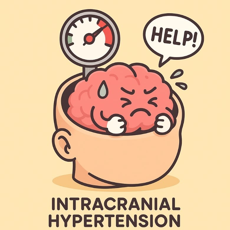
Intracranial pressure (ICP) is a frequently tested topic, both in written and oral board examinations. It is particularly relevant in patients with traumatic brain injury and during neurosurgical procedures.
Commonly tested aspects include the components and determinants of ICP, symptoms and vital signs associated with intracranial hypertension, anesthetic management, and intraoperative ICP control.

♦️ Intracranial Components
Inside the skull, there are three major constituents: brain parenchyma (~80%), cerebrospinal fluid (CSF, ~10%), and blood (~10%). (Exact ratios may differ slightly depending on the textbook.)
Since these elements are enclosed within the rigid and noncompliant skull, there is very little reserve capacity. Space-occupying lesions (tumors, hematomas) or cerebral edema can easily lead to a rise in ICP.
♦️ Normal ICP and Definition of Intracranial Hypertension
🔷 Normal ICP
These numbers must be memorized:
- Normal ICP: 5–15 mmHg
- Intervention threshold: ≥20 mmHg sustained for >5 minutes (According to the Brain Trauma Foundation [BTF] guidelines and the Society of Critical Care Medicine’s Emergency Neurological Life Support [ENLS]).
If ICP continues to rise above ~60 mmHg, global cerebral ischemia is expected, as cerebral perfusion pressure (CPP) = mean arterial pressure (MAP) – ICP.
Although some compensatory mechanisms exist (Monro–Kellie doctrine), once they are exhausted, ICP rises steeply.
🔷 Determinants of ICP
As stated abobe, ICP is determined by the pressure-volume relationships of the three intracranial components: brain tissue, CSF, and blood.
Cerebral perfusion pressure (CPP) is expressed as:
CPP = MAP – ICP
♦️ Clinical Evaluation and Symptoms of Intracranial Hypertension
It is useful to separate into subjective symptoms and objective findings.
🔷 Subjective Symptoms
Headache, nausea and vomiting, altered consciousness, seizures, and ultimately coma or respiratory arrest due to brain herniation. (Double vision may be reported, corresponding to cranial nerve VI palsy.)
🔷 Objective Findings
- Neurological signs: cranial nerve VI palsy (limitation of abduction, often reported as diplopia), papilledema.
- Vital signs: hypertension, bradycardia, and abnormal respiration — the so-called Cushing’s triad.
- Imaging: midline shift, compression of ventricles or sulci, effacement of basal cisterns, herniation in severe cases.
♦️ Anesthetic Considerations
The principle is to avoid further increases in ICP and to ensure adequate cerebral perfusion.
🔷 Induction
- Laryngoscopy, intubation, and head pin fixation can trigger sympathetic stimulation, increasing MAP and cerebral blood volume.
- Use fentanyl or remifentanil to blunt these responses.
- Avoid agents known to increase CBF and ICP such as ketamine and nitrous oxide.
- Propofol is often preferred.
- Volatile anesthetics (sevoflurane, desflurane, isoflurane) at usual clinical concentrations are generally acceptable: they dilate cerebral vessels but also reduce cerebral metabolism, resulting in balanced effects. At high concentrations, however, they may increase ICP.
- In exams and guidelines, total intravenous anesthesia (TIVA: propofol + remifentanil) is often emphasized as more favorable for ICP control.
- For rapid sequence induction (especially in emergencies): remifentanil (or fentanyl), propofol, and rocuronium are commonly used.
🔷 Maintenance
- TIVA is generally preferred.
- Avoid both hypertension (which may increase ICP at injured brain regions with impaired autoregulation) and hypotension (which may reduce CPP and cause ischemia).
- Meticulous blood pressure management is crucial.
🔷 Intraoperative ICP Control (Especially in Neurosurgery)
Since ICP is governed by brain, CSF, and blood volumes, strategies aim to reduce one or more of these components:
- Brain parenchyma:
- Surgical removal of hematomas or tumors.
- Use of mannitol (0.25–1 g/kg IV bolus) or hypertonic saline to induce osmotic diuresis.
- Loop diuretics (e.g., furosemide) may be used as adjuncts. (Note: In the U.S. and Europe, hypertonic saline has gained popularity alongside mannitol. In Japan, mannitol is still more widely used.)
- CSF volume:
- Ventricular drainage or lumbar puncture.
- (Caution: rapid CSF removal can precipitate herniation in some cases.)
- Cerebral blood volume:
- Head elevation (~30°) to facilitate venous outflow.
- Avoid neck flexion/rotation and elevated intrathoracic pressure (excessive PEEP, coughing/bucking).
- Mild hyperventilation (PaCO₂ 30–35 mmHg) can acutely reduce CBF and ICP but should be temporary, as prolonged hyperventilation increases ischemia risk.
- Correct hypoxemia, as low PaO₂ (<50–60 mmHg) markedly increases CBF and ICP.
📝 Summary(Take Home Points)
- Normal ICP: 5–15 mmHg; sustained >20 mmHg requires intervention.
- Components: brain, CSF, blood (Monro–Kellie doctrine).
- Cushing’s triad: hypertension, bradycardia, abnormal respiration.
- TIVA (propofol + remifentanil) is commonly preferred for ICP management.
- Intraoperative ICP reduction: head elevation, mild hyperventilation, mannitol/hypertonic saline, ventricular drainage, removal of mass lesions.
🔗 Related articles
- to be added
📚 References & Further reading
- Managing Intracranial Pressure Crisis(Viarasilpa,2025)
Presents comprehensive strategies for managing ICP crises, based on the most recent literature. - Monitoring the injured brain: ICP and CBF(Steiner 他,2006)
Explains the relationship between ICP and cerebral blood flow (CBF) in brain injury and the principles of monitoring. This article is highly cited. - Management of increased intracranial pressure: A review(Marik,1999)
Although relatively old, this review covers the fundamental principles of ICP management. - Initial Diagnosis and Management of Acutely Elevated ICP(Kareemi 他,2023)
Focuses on the initial evaluation and management of patients presenting with acutely elevated ICP. - Increased Intracranial Pressure(StatPearls)
Provides a concise summary of the pathophysiology, evaluation, and management strategies of ICP. - Intracranial Pressure Monitoring(StatPearls)
Outlines the techniques and clinical significance of ICP monitoring.

コメントを投稿するにはログインしてください。